an in-vitro study for early detection and to distinguish breast and lung malignancies using the pcb technology based nanodosimeter
by:Rocket PCB
2019-09-14
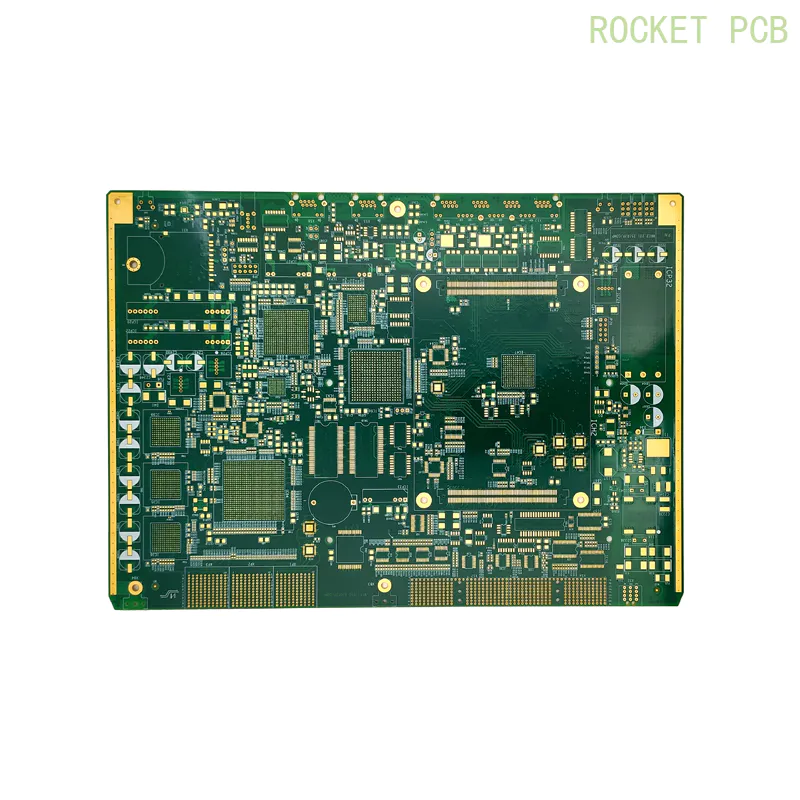
As early detection of cancer increases the chance of successful treatment, this study aims to confirm the applicability of 3D positive ion detectors based on domestic multi-layer PCB technology for early detection of breast and lung malignant tumors.
The 3D positive ion detector is a radiation detector filled with gas that works under the principle of ion-induced ionization using an exempt micro-Mrs. Curie active source.
Earlier studies reported that malignant cells could be detected by analyzing volatile organic compounds (VOCs)
Called out by cells that are outstanding biomarkers for malignant detection.
Based on this, this study analyzed the signals generated by VOCs from 140 biopsy tissue samples in the detector, which included normal tissue and all stages of breast and lung malignant tumors.
In order to strengthen the existing data, the malignant tissues of normal and advanced breast and lung were also analyzed using gas chromatography
Mass spectrometry (GC-MS).
This study confirms that the current 3D positive ion detectors can be used to detect malignant tumors in the breast and lungs, and can also distinguish them according to changes in the four basic physical parameters of the output pulse, such as frequency, amplitude, rise time and fall time, and four derived parameters of the pulse, such as FWHM, pulse area, ionization cluster size, and ion drift time.
According to the World Health Organization (WHO)
Cancer Fact 2017, cancer is the second largest cause of death (Cardio-
Vascular disease is the first cause of death in most people)
It has killed nearly six people worldwide.
About 70% of cancer deaths occur in low-and middle-income countries.
This is usually due to the growth and age of the population, lifestyle including smoking, drinking, poor diet, lack of physical activity, lack of immunity, infection, etc.
International Agency for Cancer Research (IARC)estimated 14.
There were 1 million new cases of cancer and 8 cases.
2 million cancer deaths worldwide in 2012.
WHO estimates 8.
In 2015, there were 8 million cancer deaths, including lung cancer, liver cancer, colorectal cancer, gastric cancer and breast cancer. the number of deaths was 1. 690, 0. 788, 0. 774, 0. 754, and 0.
0. 571 billion respectively.
However, IARC expects a global cancer burden of up to 21.
By 2030, there were 7 million new cancer cases and 13 million cancer deaths.
Female breast cancer and male lung cancer are the most common cancers and are also the main causes of cancer deaths for both men and women in economically developed and developing countries. in addition to lung cancer, prostate cancer is the most common cancer in men in economically developed countries.
Breast malignant tumor is the second largest cause of death in women.
It is estimated that in 2017, the 5-year survival rate of patients with breast malignant tumor was 96%, and the metastasis period was 86%, which increased to 98.
3% under localization conditions.
Therefore, early detection of breast malignant tumors is an urgent need to reduce mortality.
However, lung cancer is the fifth leading cause of death in men and women.
A total of 1 in 2012.
An estimated 8 million new lung cases, or 12.
9% of all new cancer diagnoses.
According to the Global Burden of Disease Study 2020, 5-
Annual survival rate (17. 8%)
The incidence of lung cancer is much lower than that of other major cancers.
Generally, invasive techniques such as biopsy and non-
Invasive techniques such as breast photography, ultrasound, magnetic resonance imaging (MRI)
Computed tomography (CT)
Electron emission tomography (PET)
Scanning is a common method for detecting malignant tumors in the breast and lung.
Despite the availability of effective first-line screening tools, the sensitivity of breast X-ray examination was significantly reduced in uneven and dense breast malignant tumors.
In extremely intensive cases of breast, it failed to diagnose almost half of the palpable tumors.
However, many researchers have shown that magnetic resonance and ultrasound imaging are very good methods for detecting malignant and invasive malignant tumors compared to breast X-ray examination.
These imaging strategies should be able to overcome the shortcomings of breast X-ray examination.
However, few researchers have shown that these screening methods can lead to over-diagnosis, resulting in an increase in recall rates, and a decrease in open surgical biopsy rates.
Molecular screening methods combined with these imaging techniques can help alleviate this situation.
In addition to these technologies, biomarker technology is an innovative method that has emerged over the past decade, and it can also detect malignant tumors at an early stage.
This technique also helps to monitor progress and regression after treatment of malignant tumors.
However, the biomarker technology of malignant detection involves tissues and organismsfluids.
Biomarkers may be genes, proteins, metabolites, and respiratory compounds that provide information about abnormal cell growth in malignant regions.
In order to overcome the problems that arise in the detection of these biomarkers, various technologies such as chromatography, immune tissue chemistry (IHC)
Elisa method; (ELISA)
Two-way gel swimming apparatus (2DE)
Fluorescent hybridization (FISH)
, PCR (PCR), real-
Time PCR (RT-PCR), matrix-
Laser analytical ionization time-of-flight MS (MALDI-TOF-MS), surface-
Enhanced Laser analytical Ionization Timeof-flight MS (SELDI-TOF-MS)
Malignant cells were detected by liquid chromatography combined with various detectors.
Among these technologies, gas chromatography is considered to be the most appropriate and best method due to its simple operation, selectivity and repeatability, and low detection limits.
According to the research report, GC-
MS-Based Genomics can be used to identify specific volatile organic compounds (VOCs)
As an important source biomarker for malignant diagnosis.
However, due to the lack of common metabolites, the identification or structural interpretation of these VOC molecules is limited
Specific Library
These shortcomings will be corrected by the emergence of new technologies.
Comprehensive II
Three dimensional gas chromatography (GCu2009×u2009GC)
Pair with timeof-
Flight Mass Spectrometry (TOF-MS)
The improvement of mass spectrometry instruments and identification algorithms, the development of online and open metabolic OMICS databases, etc.
Development of nanotechnology
Micro-liquid chromatography
The chip is an innovation in gas chromatography because it requires samples of nano-scale and provides nano-or low-level detection.
However, GC-
The MS technology is not flexible, slow, and requires pre-
Concentration, known requirements for the quantification of compounds, cannot be true
Time measurement, compounds should be heat stable, not suitable for clinical use, etc.
Early studies have reported that the lungs emit a variety of volatile organic compounds, including benzene, toluene, ring-based, methanol, ethanol, dicane and tripositive, the breast emission chain, olefin, ketone ester, unsaturated hydrocarbon, terpen Ene, silicone and aromatic.
In the current work, we have put forward an attempt to confirm the applicability of domestic multi-layer PCB (
Printed circuit board)
The technology-based pass 3D positive ion detector detects breast and lung malignant tumors by analyzing their signal changes, which occur due to changes in VOC emissions from malignant cells.
This detector basically combines the working principle of the thick gas electron multiplier (THGEM)
And the resistance plate counter introduced by baskirov. .
In continuing their study, we improved the efficiency of the detector by updating the detector structure, which was also recognized as a gas sensor, among other applications.
Based on the features of the 3D positive ion detector as a gas sensor, current data are obtained by collecting VOC emitted from normal and malignant breast and lung tissue, such as phase 0 (
No cancer spread), stage 1 (
Corresponding to local stage), stage 2 (
According to the participation of lymph nodes, corresponding to the local or regional stage), stage 3a (
Corresponding regional stage), stage 3b (
May have spread to lymph nodes near the chest bone)and stage 4 (
Cancer has spread to other parts of the body other than the lymph nodes)
Within a certain range of pressure (1 to 10u2009Torr).
In order to confirm the differential emission of VOCs between normal and malignant tissues, gas chromatography-
Mass spectrometry (GC-MS)
Spectrum of normal and malignant (stage 4)
Breast and lung tissue are also included.
Custom message


